
 1800 867 1390
In this week's case discussion from Dr Terry Harvey, this 20x17mm pigmented lesion is present on...

Fulfils 50 hrs for medical professionals in Australia*
100% online
Online + workshop
Fully online: $2495
Online + workshop: from $3395
Special rates available
66.5 hrs
Self-paced
2024
26 May in Melbourne
15 June in Adelaide
25 Aug is Brisbane
22 Sep in Sydney
13 Oct in Melbourne
09 Nov in Perth
 |
 |
 |


Professor and Course Coordinator MMed (Skin Cancer) Program School of Medicine, The University of Queensland, Australia
Professor Cliff Rosendahl currently works in Brisbane as a primary care practitioner with a special interest in skin cancer. He also has an interest in research as the clinical developer and Director of the Skin Cancer Audit Research Database (SCARD). His other main area of research has been in evaluating dermatoscopic clues and artificial intelligence for the diagnosis of skin malignancy in collaboration with colleagues at the Medical University of Vienna, Austria.
Prof Rosendahl has published over 70 articles in peer-reviewed journals and authored/co- authored two textbooks. He has a busy schedule presenting to GPs in Australia and to GPs and dermatologists internationally.

Dr Colin Armstrong is a general practitioner and part-time clinical trials investigator at the Wesley Research Institute, Brisbane. He completed all HealthCert certificate courses and The University of Queensland Master of Medicine (Skin Cancer) in 2011. Working primarily in skin cancer since 2010, Colin is passionate about building GPs’ confidence in their management of skin cancer and has an ongoing commitment to GP education in all facets of skin cancer diagnosis and treatment.

Doctor, National Skin Cancer Centres, Berwick
Dr Hamilton Ayres worked in Adelaide as a Plastic Surgery Registrar at Flinders, Repatriation General Hospital and the Royal Adelaide Hospital where his main role was the management of trauma, hand injuries and difficult skin cancers. Hamilton has obtained a Fellowship of the Royal Australian College of General Practitioners and Certificates in Skin Cancer Medicine, Dermatoscopy and Histopathology from HealthCert and The University of Queensland School of Medicine.


In this week's case discussion from Dr Terry Harvey, this 20x17mm pigmented lesion is present on...
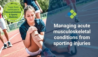
Acute musculoskeletal injuries acquired during sport are a serious concern. These are medical...
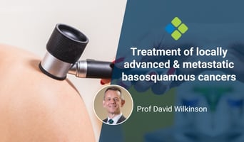
This month we touch on an uncommon but important topic – that of locally advanced and metastatic...


