
 1800 867 1390
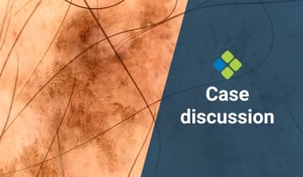
In this week's case discussion from Dr Terry Harvey, this 20x17mm pigmented lesion is present on...

Fulfils 50 hrs for medical professionals in Australia
100% online
$1995
Special rates available
80 hrs
Self-paced
 |
 |
 |

Professor at the Department of Dermatology, Medical University of Vienna, Austria
Professor Harald Kittler has a special clinical interest in dermoscopy of pigmented skin lesions. His main research interest is digital dermoscopy, follow-up of pigmented skin lesions, and computer assisted digital dermoscopy. Harald has been working for 10 years in the field of dermoscopy and has published a number of scientific articles especially in the field of digital dermoscopy and dermoscopic follow-up of melanocytic nevi.

Dermatologist-Venereologist, First Department Of Dermatology, Aristotle University, Greece
President, International Dermoscopy Society
Aimilios Lallas is an Associate Professor of Dermatology at the First Department of Dermatology of Aristotle University in Thessaloniki, Greece. He is specialised in skin cancer diagnosis with non-invasive techniques, as well as in the management of skin cancer patients.
His main field of research interest is dermoscopy of skin tumours, the application of the method in general dermatology and the improvement of the management of oncologic patients. He is an author of more than 330 scientific papers published on Pubmed Central, most of them on dermoscopy and skin cancer. He is an editor of eight books and author of several book chapters on dermoscopy. He is a co-investigator in several Phase III Clinical trials on skin cancer treatment. He has been awarded several scholarships and scientific awards.
Over the last years, A/Prof Lallas has established scientific collaboration with numerous colleagues from several countries and has supervised the training of numerous fellows from different countries. He is an invited speaker in several domestic and international congresses and meetings, mainly on dermoscopy and on skin cancer diagnosis and management. He is particularly involved in teaching activities on dermoscopy, having organised and participated in numerous domestic and international courses.
A/Prof Lallas is currently the President of the International Dermoscopy Society.

Scientific Coordinator, Skin Cancer Unit, ASMN-IRCCS, University of Modena and Reggio Emilia, Italy
Associate Professor Caterina Longo is a board-certified dermatologist specialising in the diagnosis and treatment of skin cancers. Although providing the best care possible for patients remains her primary goal, she also committed to education and clinical research. She is actively involved in clinical research and has published numerous papers on topics related to skin cancer with an emphasis on melanoma, atypical nevi, Spitz/Reed nevi and non-melanoma skin cancer.
Caterina’s research interests are focused on the use of imaging instruments such as dermoscopy and confocal laser microscopy to recognise skin cancer early in its development. She pioneered the use of ex vivo fluorescence confocal microscopy for micrographic Mohs surgery applied for basal cell carcinoma and other visceral tumours. Caterina lectures on these topics both nationally and internationally.

Head of the Dermatology Clinic of the University of Trieste, Italy
Associate Professor Iris Zalaudek is a board-certified dermatologist and Head of the Dermatology Clinic of the University of Trieste, Italy. Since 2016, she has been President of the International Dermoscopy Society, and was previously the Research Director of the Non-Melanoma Skin Cancer Unit at the Medical University of Graz, Austria.
Her main research fields are related to dermato-oncology and include non-invasive skin imaging techniques, as well as topical and systemic treatment of skin cancer. Moreover, she is engaged in the development of modern teaching methods such as online distant courses and tele-dermatologic services. She is Director of the Master of Science program entitled "Dermoscopy and Preventive Dermato-Oncology" of the Medical University of Graz, Austria.
Iris has published more than 450 articles, of which 358 (267 full papers) have been cited in PubMed. Her combined publications have received an impact factor of 1003 and a h-index value of 36 (by April 2017). In 2003 her work was awarded by the Hans-Weitgasser Price from the Styrian Association of Dermatologists and in 2008 she was awarded the Best Researcher of the Medical University of Graz, Austria.

Dermatologist, Santa Maria Nuova Hospital, Reggio Emilia, Italy
Dr Elvira Moscarella is a dermatologist at the Santa Maria Nuova Hospital in Reggio Emilia, Italy. She acquired her medical degree in 2005 at the Second University of Naples before completing her residency in dermatology and venereology at the University’s Department of Dermatology. In 2008, Elvira undertook further education in dermoscopy and confocal microscopy. She is a member of the European Academy of Dermatology and Venereology and the International Dermoscopy Society, and is Editor in Chief of the latter’s newsletter and case of the month. Elvira’s main interests are in dermoscopy and reflectance confocal microscopy, and their use in skin cancer medicine.


In this week's case discussion from Dr Terry Harvey, this 20x17mm pigmented lesion is present on...

Acute musculoskeletal injuries acquired during sport are a serious concern. These are medical...
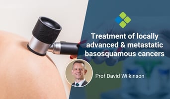
This month we touch on an uncommon but important topic – that of locally advanced and metastatic...


