
 1800 867 1390
In this week's case discussion from Dr Terry Harvey, this 20x17mm pigmented lesion is present on...


Fulfils 50 hrs for medical professionals in Australia*
100% online
$1995
Special rates available
81.5 hrs
Self-paced


Honorary Lecturer in Pathology, The University of Queensland
Dr Simon Clark runs the pathology component of the postgraduate certificate courses in skin cancer medicine at The University of Queensland and lectures in the Masters of Medicine course. He has been involved in dermatopathology education for more than 20 years, training registrars in dermatology, pathology and plastic surgery. More recently he has been active in GP education. One of the best known dermatopathologists in Australia, Simon was recently appointed a visiting professor in dermatology at the Tehran University of Medical Sciences.


In this week's case discussion from Dr Terry Harvey, this 20x17mm pigmented lesion is present on...

Acute musculoskeletal injuries acquired during sport are a serious concern. These are medical...
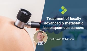
This month we touch on an uncommon but important topic – that of locally advanced and metastatic...


